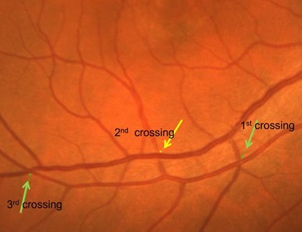
Hypothetical proposal for the course of retinal blood vessels in the posterior pole—description and its clinical implications
Medical hypothesis, discovery & innovation in optometry,
Vol. 4 No. 4 (2023),
25 December 2023
,
Page 188-194
https://doi.org/10.51329/mehdioptometry190
Abstract
Background: Branch retinal vein occlusion (BRVO) is the second-most common retinal vascular disorder. Arteriosclerotic changes at the site of obstruction and hemodynamic turbulence within the vessels are considered risk factors. Overcrossing of the vein by an artery has traditionally been considered to increase the risk of BRVO. Recent studies using optical coherence tomography and optical coherence tomography angiography have suggested a higher prevalence of vein-over-artery crossings in this disease. Nevertheless, uncertainty persists as to why some patients, even those with the same disease duration, have varying degrees of venous dilation and develop sufficient collaterals, while others develop substantial ischemia and its sequelae.Hypothesis: Herein, it is hypothesized that because retinal blood vessels are transparent, tubular, and collapsible conduits coursing over a hollow spherical surface, the changes related to AV crossings over the entire course of a vessel, rather than at any single isolated crossing, could contribute to the risk, natural progression, and outcomes of BRVO. The study analyzed color fundus photographs from two image datasets. The first dataset comprised 100 randomly selected images from the author’s own collection at the Rajendra Prasad Center for Ophthalmic Sciences. The second dataset comprised 100 images from the MESSIDOR database; three images were excluded owing to poor focus. Using 394 observations from 197 retinal photographs, four distinct patterns of AV crossing along the course of blood vessels were recognized: (A and B) wicker basket, (C) straight, (D) widely spaced, and (E) indeterminate. The percentages of tight wicker, loose wicker, straight, widely spaced, and indeterminate patterns in the two image sets were 19% (38/200) and 16.5% (32/194), 22.5% (45/200) and 27.8% (54/194), 16.5% (33/200) and 15.5% (30/194), 22.5% (45/200) and 28.4% (55/194), and 19.5% (39/200) and 11.9% (23/194), respectively. Hence, the wicker basket pattern was the most common AV crossing pattern in both image sets.
Conclusions: The wicker basket pattern may provide structural stability and aid in maintaining pressure gradients within the retinal vascular bed. This observation of variable AV relationships at consecutive crossings may improve our understanding of the pathogenesis, natural history, and outcomes of BRVO. Future longitudinal studies including patients at risk of BRVO, or retrospective analyses of patients with BRVO who had ophthalmic examinations and archived fundus images before the vascular event, should verify the relevance of these observed vascular patterns.
Keywords:
- blood vessel
- retinal vessel
- branch retinal vein occlusion
- hypothesis
- observation

References
1. Ramos D, Catita J, López-Luppo M, Valença A, Bonet A, Carretero A, et al. Vascular Interstitial Cells in Retinal Arteriolar Annuli Are Altered During Hypertension. Invest Ophthalmol Vis Sci. 2019;60(2):473-487. doi: 10.1167/iovs.18-25000 pmid: 30707220
2. Fukutsu K, Saito M, Noda K, Murata M, Kase S, Shiba R, et al. A Deep Learning Architecture for Vascular Area Measurement in Fundus Images. Ophthalmol Sci. 2021;1(1):100004. doi: 10.1016/j.xops.2021.100004 pmid: 36246007
3. Wong TY, Klein R, Klein BE, Meuer SM, Hubbard LD. Retinal vessel diameters and their associations with age and blood pressure. Invest Ophthalmol Vis Sci. 2003;44(11):4644-50. doi: 10.1167/iovs.03-0079 pmid: 14578380
4. Miri M, Amini Z, Rabbani H, Kafieh R. A Comprehensive Study of Retinal Vessel Classification Methods in Fundus Images. J Med Signals Sens. 2017;7(2):59-70 pmid: 28553578
5. Wong TY, Klein R, Klein BE, Tielsch JM, Hubbard L, Nieto FJ. Retinal microvascular abnormalities and their relationship with hypertension, cardiovascular disease, and mortality. Surv Ophthalmol. 2001;46(1):59-80. doi: 10.1016/s0039-6257(01)00234-x pmid: 11525792
6. Hayreh SS. Ocular vascular occlusive disorders: natural history of visual outcome. Prog Retin Eye Res. 2014;41:1-25. doi: 10.1016/j.preteyeres.2014.04.001 pmid: 24769221
7. Hirano Y, Suzuki N, Tomiyasu T, Kurobe R, Yasuda Y, Esaki Y, et al. Multimodal Imaging of Microvascular Abnormalities in Retinal Vein Occlusion. J Clin Med. 2021;10(3):405. doi: 10.3390/jcm10030405 pmid: 33494354
8. Uhl A, Busch C, Marcel S, Veldhuis R (2020). ‘Handbook of vascular biometrics’. (pp. 1-533).Springer Nature. doi: 10.1007/978-3-030- 27731-4
9. Iida Y, Muraoka Y, Ooto S, Suzuma K, Murakami T, Iida-Miwa Y, et al. Morphologic and Functional Retinal Vessel Changes in Branch Retinal Vein Occlusion: An Optical Coherence Tomography Angiography Study. Am J Ophthalmol. 2017;182:168-179. doi: 10.1016/j.ajo.2017.08.004 pmid: 28837791
10. Anand-Apte B, Hollyfield JG (2010). ‘Developmental Anatomy of the Retinal and Choroidal Vasculature’. (pp. 1-15).Elsevier Ltd. Link
11. MESSIDOR (2004). ‘Methods for Evaluating Segmentation and Indexing techniques Dedicated to Retinal Ophthalmology’. MESSIDOR is a project funded by the French Ministry of Research and Defense within a 2004 TECHNO-VISION program. Available at: https://www.adcis.net/en/third-party/messidor/ (Accessed: April 22, 2020)
12. Decencière E, Zhang X, Cazuguel G, Lay B, Cochener B, Trone C, et al. Feedback on a publicly distributed image database: the Messidor database. Image Analysis & Stereology. 2014; 33(3):231-4. doi: 10.5566/ias.1155
13. Joyal JS, Gantner ML, Smith LEH. Retinal energy demands control vascular supply of the retina in development and disease: The role of neuronal lipid and glucose metabolism. Prog Retin Eye Res. 2018;64:131-156. doi: 10.1016/j.preteyeres.2017.11.002 pmid: 29175509
14. Sun C, Chen T, Cong J, Wu X, Wang J, Yuan Y. Changes in retinal vascular bifurcation in eyes with myopia. BMC Ophthalmol. 2022;22(1):408. doi: 10.1186/s12886-022-02629-y pmid: 36271390
15. Spaide RF, Gemmy Cheung CM, Matsumoto H, Kishi S, Boon CJF, van Dijk EHC, et al. Venous overload choroidopathy: A hypothetical framework for central serous chorioretinopathy and allied disorders. Prog Retin Eye Res. 2022;86:100973. doi: 10.1016/j. preteyeres.2021.100973 pmid: 34029721
16. Stefánsson E, Chan YK, Bek T, Hardarson SH, Wong D, Wilson DI. Laws of physics help explain capillary non-perfusion in diabetic retinopathy. Eye (Lond). 2018;32(2):210-212. doi: 10.1038/eye.2017.313 pmid: 29350688
17. Takahashi K, Song Y, Sogawa K, Yoshioka T, Tani T, Ishiko S, et al. Deterioration of Retinal Blood Flow Parameters in Branch Retinal Vein Occlusion Measured by Doppler Optical Coherence Tomography Flowmeter. J Clin Med. 2020;9(6):1847. doi: 10.3390/ jcm9061847 pmid: 32545765
18. Rehak J, Rehak M. Branch retinal vein occlusion: pathogenesis, visual prognosis, and treatment modalities. Curr Eye Res. 2008;33(2):111- 31. doi: 10.1080/02713680701851902 pmid: 18293182
19. Laouri M, Chen E, Looman M, Gallagher M. The burden of disease of retinal vein occlusion: review of the literature. Eye (Lond). 2011;25(8):981-8. doi: 10.1038/eye.2011.92 pmid: 21546916
20. Khoo YJ, Yu DY, Abdul-Rahman A, Balaratnasingam C, Chen FK, McAllister IL, et al. Vessel Pulse Amplitude Mapping in Eyes With Central and Hemi Retinal Venous Occlusion. Transl Vis Sci Technol. 2023;12(1):26. doi: 10.1167/tvst.12.1.26 pmid: 36692455
21. Tomita R, Goto K, Ueno Y, Yamaguchi K, Takeuchi J, Akahori T, et al. Narrowing Ratio of Retinal Veins at Arteriovenous Crossing in Patients With Branch Retinal Vein Occlusion Versus That in Healthy Individuals. Invest Ophthalmol Vis Sci. 2023;64(14):22. doi:
10.1167/iovs.64.14.22 pmid: 37971732 22. Jaulim A, Ahmed B, Khanam T, Chatziralli IP. Branch retinal vein occlusion: epidemiology, pathogenesis, risk factors, clinical features, diagnosis, and complications. An update of the literature. Retina. 2013;33(5):901-10. doi: 10.1097/IAE.0b013e3182870c15 pmid:
23609064
23. Darabu? DM, Munteanu M, Preda MA, Karancsi OL, ?u?? MC. The Impact of Intraocular Treatment on Visual Acuity of Patients Diagnosed with Branch Retinal Vein Occlusions. Healthcare (Basel). 2023;11(10):1414. doi: 10.3390/healthcare11101414 pmid: 37239696
24. Muraoka Y, Tsujikawa A. Arteriovenous crossing associated with branch retinal vein occlusion. Jpn J Ophthalmol. 2019;63(5):353-364. doi: 10.1007/s10384-019-00676-5 pmid: 31396750
25. Kumagai K, Tsujikawa A, Muraoka Y, Akagi-Kurashige Y, Murakami T, Miyamoto K, et al. Three-dimensional optical coherence tomography evaluation of vascular changes at arteriovenous crossings. Invest Ophthalmol Vis Sci. 2014;55(3):1867-75. doi: 10.1167/ iovs.13-13303 pmid: 24576872
26. Parodi MB, Bandello F. Branch retinal vein occlusion: classification and treatment. Ophthalmologica. 2009;223(5):298-305. doi: 10.1159/000213640 pmid: 19372724
27. Rehak M, Wiedemann P. Retinal vein thrombosis: pathogenesis and management. J Thromb Haemost. 2010;8(9):1886-94. doi: 10.1111/j.1538-7836.2010.03909.x pmid: 20492457
28. Muraoka Y, Tsujikawa A, Murakami T, Ogino K, Kumagai K, Miyamoto K, et al. Morphologic and functional changes in retinal vessels associated with branch retinal vein occlusion. Ophthalmology. 2013;120(1):91-9. doi: 10.1016/j.ophtha.2012.06.054 pmid: 22980743
29. Kogo T, Muraoka Y, Iida Y, Ooto S, Murakami T, Kadomoto S, et al. Angiographic Risk Features of Branch Retinal Vein Occlusion Onset as Determined by Optical Coherence Tomography Angiography. Invest Ophthalmol Vis Sci. 2020;61(2):8. doi: 10.1167/ iovs.61.2.8 pmid: 32031580
- Abstract Viewed: 0 times
- Full Text PDF Downloaded: 0 times