IVORC
  • Register
  • Login

Medical hypothesis, discovery & innovation in optometry

  1. Home
  2. Archives
  3. Vol. 3 No. 4 (2022): Winter 2022
  4. Articles

About the Journal

Editorial Team

Privacy Statement

Contact

Vortex keratopathy after photorefractive keratectomy in a patient on long-term clozapine treatment

  • Samir Shoughy
  • Hosam Elgemie
  • Waleed Nasif

Medical hypothesis, discovery & innovation in optometry, Vol. 3 No. 4 (2022), 14 January 2023 , Page 160-164
https://doi.org/10.51329/mehdioptometry165 Published 14 January 2023

  • View Article
  • Download
  • References
  • Share

Abstract

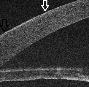
Background: Vortex keratopathy is a corneal entity characterized by corneal deposits at the level of the basal epithelium in the form of a whorl-like pattern in the interpalpebral portions of the cornea. Medications such as amiodarone, chloroquine, hydroxychloroquine, indomethacin, phenothiazines, ibuprofen, and naproxen bind to the cellular lipids of the basal epithelial layer of the cornea, producing a characteristic whorl pattern. Here, we report a case of bilateral central vortex keratopathy with slight vision impairment 3 weeks after uneventful photorefractive keratectomy (PRK) in a woman on long-term clozapine treatment.
Case Presentation: A 42-year-old woman presented to the outpatient clinic for refractive surgery for the correction of bilateral moderate myopia. Her best-corrected distance visual acuity was 20 / 20 in both eyes. She had been on antipsychotic clozapine tablets 400 mg / day for the past 6 years to manage a psychiatric disorder. On detailed preoperative slit-lamp examination, her corneas were clear, with perilimbal conjunctival pigmentations. Other ocular examination results were unremarkable. At the 3-week follow-up after an uneventful PRK, her uncorrected distance visual acuity was 20 / 25 in the right eye and 20 / 20 in the left eye, with complaints of slightly unclear vision in both eyes. Slit-lamp examination revealed bilateral corneal subepithelial deposits in a whorl-like pattern reminiscent of vortex keratopathy. The deposits were restricted to the epithelial and subepithelial regions and did not extend to the stroma without evident stromal haze. Dilated fundus examination results were normal bilaterally. Despite excellent refractive outcomes and visual acuity in both eyes, she was dissatisfied and complained of unclear vision. She was administered with lubricating eye drops and placed on regular follow-ups. At the 4-month follow-up, the vortex lines were cleared centrally, and she reported improvement in vision. Anterior-segment optical coherence tomography of the cornea revealed no epithelial deposits in the central part of the cornea and residual deposits in the temporal peripheral cornea, indicating clearing of the deposits from the central cornea and explaining the subjective improvement in vision. Her final best-corrected distance visual acuity was 20/20 in both eyes with an unremarkable ocular examination.
Conclusions: Our case indicates a potential causal relationship between long-term clozapine treatment and development of temporary, visually significant vortex keratopathy after uneventful PRK. Further large-scale studies are required to verify the causal relationship between the long-term clozapine administration and the development of vortex keratopathy following surface ablation photorefractive procedures.
Keywords:
  • photorefractive keratectomies
  • visual acuities
  • vortex keratopathy
  • optical coherence tomography
  • clozaril
  • side effects
  • corneas
  • conjunctivas
  • Full Text PDF

References

Raizman MB, Hamrah P, Holland EJ, Kim T, Mah FS, Rapuano CJ, et al. Drug-induced corneal epithelial changes. Surv Ophthalmol. 2017;62(3):286-301. doi: 10.1016/j.survophthal.2016.11.008 pmid: 27890620

Sahyoun JY, Sabeti S, Robert MC. Drug-induced corneal deposits: an up-to-date review. BMJ Open Ophthalmol. 2022;7(1):e000943. doi: 10.1136/bmjophth-2021-000943 pmid: 35415268

Hollander DA, Aldave AJ. Drug-induced corneal complications. Curr Opin Ophthalmol. 2004;15(6):541-8. doi: 10.1097/01.icu.0000143688.45232.15 pmid: 15523201

Gitahy Falcao Faria C, Weiner L, Petrignet J, Hingray C, Ruiz De Pellon Santamaria Á, Villoutreix BO, et al. Antihistamine and cationic amphiphilic drugs, old molecules as new tools against the COVID-19? Med Hypotheses. 2021;148:110508. doi: 10.1016/j.mehy.2021.110508 pmid: 33571758

Wolf K, Kühn H, Boehm F, Gebhardt L, Glaudo M, Agelopoulos K, et al. A group of cationic amphiphilic drugs activates MRGPRX2 and induces scratching behavior in mice. J Allergy Clin Immunol. 2021;148(2):506-522.e8. doi: 10.1016/j.jaci.2020.12.655 pmid: 33617860

Wu MK, Chung W, Wu CK, Tseng PT. The severe complication of Stevens-Johnson syndrome induced by long-term clozapine treatment in a male schizophrenia patient: a case report. Neuropsychiatr Dis Treat. 2015;11:1039-41. doi: 10.2147/NDT.S79327 pmid: 25914536

Borovik AM, Bosch MM, Watson SL. Ocular pigmentation associated with clozapine. Med J Aust. 2009;190(4):210-1. doi: 10.5694/j.1326-5377.2009.tb02353.x pmid: 19220190

Ceylan E, Ozer MD, Yilmaz YC, Kartal B, Yildiz Ekinci D, Çinici E, et al. The ocular surface side effects of an anti-psychotic drug, clozapine. Cutan Ocul Toxicol. 2016;35(1):62-6. doi: 10.3109/15569527.2015.1018387 pmid: 25853177

Alam MS, Praveen Kumar KV. Clozapine-induced cataract in a young female. J Pharmacol Pharmacother. 2016;7(4):184-186. doi: 10.4103/0976-500X.195904 pmid: 28163541

Moshirfar M, Bundogji N, Tukan AN, Ronquillo YC. Implications of Corneal Refractive Surgery in Patients with Fabry Disease. Ophthalmol Ther. 2022;11(3):925-929. doi: 10.1007/s40123-022-00503-0 pmid: 35438438

Lim L, Wei RH. Laser in situ keratomileusis treatment for myopia in a patient with partial limbal stem cell deficiency. Eye Contact Lens. 2005;31(2):67-9. doi: 10.1097/01.icl.0000146302.00152.47 pmid: 15798476

Schroeder FM, Hörle S. Zentrale Cornea verticillata nach Lasik bei Amiodaron-Therapie [Vortex keratopathy with unusual central position after LASIK-treatment with amiodarone]. Klin Monbl Augenheilkd. 2009;226(1):66-7. German. doi: 10.1055/s-2008-1027841 pmid: 19173166

Karampatakis V, Karamitsos A, Skriapa A, Pastiadis G. Comparison between normal values of 2- and 5-minute Schirmer test without anesthesia. Cornea. 2010;29(5):497-501. doi: 10.1097/ICO.0b013e3181c2964c pmid: 20299972

El-Sersy TH. Assessment of posterior corneal surface changes after photorefractive keratectomy in moderate myopia. Journal of the Egyptian Ophthalmological Society. 2016;109(4):167-171. doi: 10.4103/2090-0686.204728

Turk U, Turk BG, Y?lmaz SG, Tuncer E, Alio?lu E, Dereli T. Amiodarone-induced multiorgan toxicity with ocular findings on confocal microscopy. Middle East Afr J Ophthalmol. 2015;22(2):258-60. doi: 10.4103/0974-9233.154411 pmid: 25949090

Tomás-Juan J, Murueta-Goyena Larrañaga A, Hanneken L. Corneal Regeneration After Photorefractive Keratectomy: A Review. J Optom. 2015;8(3):149-69. doi: 10.1016/j.optom.2014.09.001 pmid: 25444646

Ikegawa Y, Shiraishi A, Hayashi Y, Ogimoto A, Ohashi Y. In Vivo Confocal Microscopic Observations of Vortex Keratopathy in Patients with Amiodarone-Induced Keratopathy and Fabry Disease. J Ophthalmol. 2018;2018:5315137. doi: 10.1155/2018/5315137 pmid: 29750121

Nejima R, Miyata K, Tanabe T, Okamoto F, Hiraoka T, Kiuchi T, et al. Corneal barrier function, tear film stability, and corneal sensation after photorefractive keratectomy and laser in situ keratomileusis. Am J Ophthalmol. 2005;139(1):64-71. doi: 10.1016/j.ajo.2004.08.039 pmid: 15652829

Jung JW, Kim JY, Chin HS, Suh YJ, Kim TI, Seo KY. Assessment of meibomian glands and tear film in post-refractive surgery patients. Clin Exp Ophthalmol. 2017;45(9):857-866. doi: 10.1111/ceo.12993 pmid: 28544605

Rosa N, Borrelli M, De Bernardo M, Lanza M. Corneal morphological changes after myopic excimer laser refractive surgery. Cornea. 2011;30(2):130-5. doi: 10.1097/ICO.0b013e3181f237a1 pmid: 21045644

Ortega-Usobiaga J, Llovet-Osuna F, Reza Djodeyre M, Cobo-Soriano R, Llovet-Rausell A, Baviera-Sabater J. LASIK and surface ablation in patients treated with amiodarone. Arch Soc p Oftalmol. 2016;91(11):520-525. English, Spanish. doi: 10.1016/j.oftal.2016.05.005 pmid: 27350387

Reynolds A, Moore JE, Naroo SA, Moore CB, Shah S. Excimer laser surface ablation - a review. Clin Exp Ophthalmol. 2010;38(2):168-82. doi: 10.1111/j.1442-9071.2010.02230.x pmid: 20398106

Jain KK (2021). ‘Drug-induced neurological disorders’. Springer Nature; 4th edn (pp. 413-496). Publisher: Springer Cham. eBook ISBN: 978-3-030-73503-6. doi: 10.1007/978-3-030-73503-6

Kushner BJ, Kowal L. Diplopia after refractive surgery: occurrence and prevention. Arch Ophthalmol. 2003;121(3):315-21. doi: 10.1001/archopht.121.3.315 pmid: 12617699

Finlay AL. Binocular vision and refractive surgery. Cont Lens Anterior Eye. 2007;30(2):76-83. doi: 10.1016/j.clae.2007.02.009 pmid: 17448926

García-Montero M, Albarrán Diego C, Garzón-Jiménez N, Pérez-Cambrodí RJ, López-Artero E, Ondategui-Parra JC. Binocular vision alterations after refractive and cataract surgery: a review. Acta Ophthalmol. 2019;97(2):e145-e155. doi: 10.1111/aos.13891 pmid: 30218490

  • Abstract Viewed: 0 times
  • Full Text PDF Downloaded: 0 times

Download Statastics

  • Linkedin
  • Twitter
  • Facebook
  • Google Plus
  • Telegram
Make a Submission
Information
  • For Readers
  • For Authors
  • For Librarians
  • Home
  • Archives
  • Submissions
  • About the Journal
  • Editorial Team
  • Contact

This work is licensed under a Creative Commons Attribution-NoDerivatives 4.0 International License.

© Copyright 2021-2022 CC BY-NC 4.0. All Rights Reserved.

Medical Hypothesis, Discovery & Innovation in Optometry
ISSN 2693-8391