Evaluating the predictive value of optical coherence tomography angiography metrics and central corneal thickness among glaucoma suspect patients: a comparative cross-sectional study
Medical hypothesis discovery and innovation in ophthalmology,
Vol. 14 No. 4 (2025),
17 December 2025
,
Page 183-193
Abstract
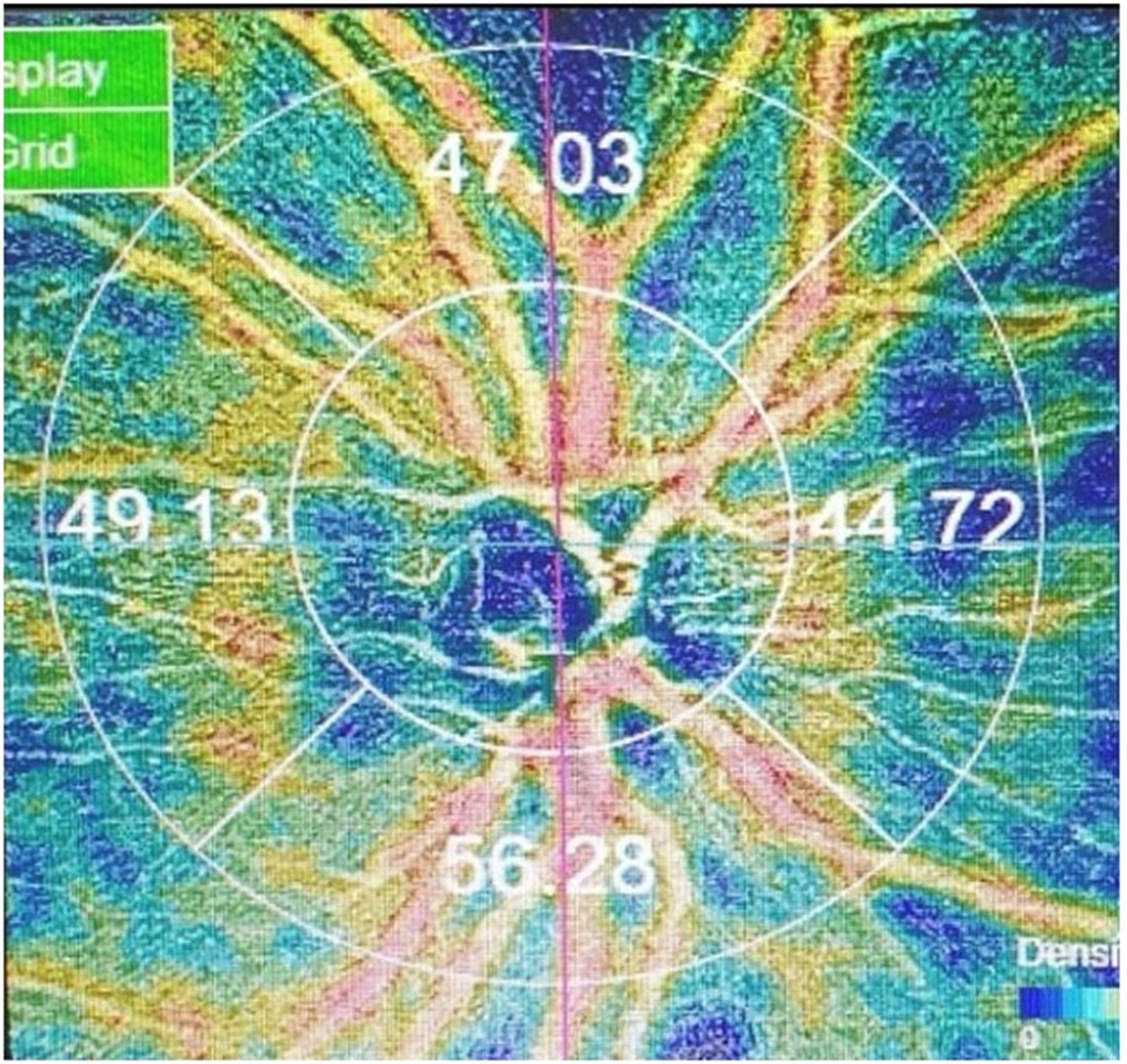
Background: Glaucoma suspects (GS) exhibit risk factors such as elevated intraocular pressure (IOP), suspicious optic disc or retinal nerve fiber layer (RNFL) findings, or a positive family history, yet their risk of progression varies widely. Optical coherence tomography angiography (OCTA) and central corneal thickness (CCT) have emerged as important markers for detecting early structural and microvascular changes in GS patients. We aimed to estimate the predictive value of OCTA-derived metrics and CCT, and to assess their correlations in GS individuals.Methods: This comparative cross-sectional study included eyes from GS patients and eyes from age- and sex-matched healthy individuals as a comparison group. All participants underwent a detailed medical history and comprehensive ophthalmologic examination. Investigations included visual field perimetry; optical coherence tomography (OCT) to assess structural optic nerve head parameters, RNFL thickness, and ganglion cell layer (GCL) thickness; OCTA to measure papillary vascular density (PVD) and radial peripapillary capillary density (RPC); and non-contact specular microscopy to determine CCT.
Results: The GS group had a mean age of 36.9 years, with 52.4% male (n = 11). GS eyes showed significantly larger CDR values, reduced rim area, thinner RNFL and GCL, and lower CCT compared with healthy eyes (all P < 0.05). Mean RPC, quadrant-specific RPC values, and mean PVD were significantly reduced in GS individuals (all P < 0.05). CCT showed significant correlations with all vascular metrics and structural parameters (all P < 0.05), except disc area (P > 0.05). In univariate logistic regression all variables were associated with GS status. After multivariate adjustment, only CCT less than or equal to 506 µm remained an independent predictor. Receiver operating characteristic curve analysis showed good diagnostic performance for CCT (area under the curve [AUC] = 0.757) and mean RPC (AUC = 0.820) in identifying GS eyes.
Conclusions: Patients with GS revealed significantly lower structural parameters and vascular metrics compared with the healthy group, and only thin CCT remained an independent predictor of GS status. Both CCT and mean RPC demonstrated good diagnostic performance for identifying GS eyes.
- Abstract Viewed: 0 times
- Full Text PDF Downloaded: 0 times
